

Treatment of traumatic peripheral nerve injuries in the extremities

OPERATIONS
Shoulder arthroscopy Hip endoprosthetics Bunion correction surgery Stem cells ACL reconstruction Knee endoprosthetics Brachial plexus reconstruction Treatment of traumatic peripheral nerve injuries in the extremities Surgical treatment of neoplastic and tumour-like lesions of the peripheral nerves and brachial plexus High density platelet-rich plasma Carpal Tunel release procedure Knee Arthroscopy Hip ArthroscopyOPERATIONS
Traumatic nerve damage can be open or closed. The former is the result of injuries (cuts, lacerations, puncture wounds, gunshot wounds), in which the continuity of the integumentary system is interrupted with accompanied damage to nerve structures and possibly neighbouring vessels. This mechanism of injury carries a high risk of complete interruption of nerve continuity.
Closed injuries are caused by the stretching of the nerve (so-called traction injuries), accompanied by secondary compression associated with gradual fibrosis of the hematoma. Stretching of the nerve can be caused by a displaced bone fragment in a fracture or an articular surface in a dislocation. Nerve traction damage can also occur as a result of sudden stretching or jerking of the limb. Closed injuries can also be caused by blunt force trauma (contusions), accompanied by the formation of a hematoma in the nerve area.
Nerve injuries can have varying degrees of severity, which are graded on Sunderland’s five-grade scale. Grade I and II have favourable prognoses, and in these cases, it is possible to recover nerve function with conservative treatment. In grade V, where all nerve components are subject to complete interruption, recovery of function is possible only as a result of surgical treatment (microsurgical reconstruction).
In the case of damage to mixed nerves, consisting of motor, sensory and autonomic fibres, symptoms will be associated with impaired function of all these structures. The patient will experience sensory disturbances in the innervation area of the nerve in question, as well as muscle atrophy with inability to perform movements in the joints. Irritation of the autonomic system will cause vasomotor and secretory disorders, pain, and in extreme cases ulceration. Damage to individual nerves results in certain characteristic clinical symptoms.
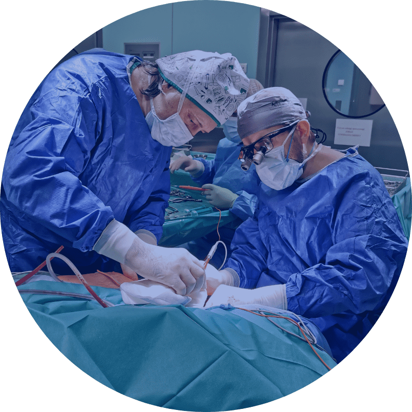
Various microsurgical techniques, such as neurolysis, reconstruction with direct suture, reconstruction with cutaneous nerve grafts (possibly neurotubes or allografts), and extra-anatomical reconstruction are possible during the surgery.
Neurolysis involves freeing the nerve from adhesions. It is a technique used in cases where the preserved nerve continuity and its compression by scar tissue is found intraoperatively.
Nerve continuity interruption requires microsurgical reconstruction. For small defects, it is possible to achieve union of both nerve stumps with a direct suture. Larger cavities of several centimetres require grafting. Usually the sural nerve (sensory nerve) taken from the lower leg is used for this purpose.
An alternative to using sensory nerves is to use neurotubes or allografts. Neurotubes are cylindrical structures into which nerve stumps are inserted. Neurotubes made of collagen, chitosan, polyglycolic acid and a combination of polylactic acid and caprolactone are commercially available. Allografts are fragments of nerves taken from deceased donors, which are devoid of their antigenic properties as a result of heat, enzymatic and radiation treatment. Neurotubes and allografts allow replacing small defects in relatively thin nerves, such as finger nerves. When reconstructing them, it is also possible to use a section of a vein.
Extra-anatomical reconstruction involves restoring the function of an important nerve by “sacrificing” the function of another, less clinically important, nerve.
Nerve unions are created using very thin sutures and fibrin glue. The procedures are performed either under a microscope or with the use of surgical magnifying loupes.
The surgical outcomes depend on the patient’s age, time from injury to surgery, the size of the nerve defect, and the level and degree of damage.
The nerve regenerative potential is the greatest in young and middle-aged people. Above the age of 65, it becomes weaker. This does not rule out the chance of improvement in older people, but it is usually lower.
The best outcomes of surgical treatment of traumatic peripheral nerve injuries are obtained when the nerve is managed immediately after the injury (primary suture or delayed primary suture) or after secondary reconstructions performed up to 6 months after the injury. Prolonged denervation of the muscles causes their gradual, with time irreversible, atrophy. Degenerative changes also occur in the neuromuscular junctions. After 18 months from the injury, these changes become irreversible, and reconstruction of the nerve in these cases is unwarranted (so-called old nerve damage).
A large defect in the continuity of the nerve is a prognostically unfavourable factor. Its size depends, of course, on the mechanism of injury, but also on the timing of surgical treatment. Over time, neurofibromas develop in the stumps of the interrupted nerve (the larger one in the proximal part of the limb). During reconstruction, it is necessary to excise them, which increases the extent of the defect.
High nerve damage (located in the proximal parts of the limb) has a worse prognosis than more peripheral damage. This is related to the longer regeneration path of axons and their constant, relatively slow, growth rate (1 mm/day).
Preoperative instructions:
Diagnosis of traumatic nerve injuries includes clinical examination and adjunct tests (electromyography, imaging tests: ultrasound and MRI).
Postoperative instructions:
Ensure proper wound healing, maintain immobilization of the limb as recommended, then start rehabilitation, muscle stimulation – 6 weeks after reconstruction, avoid cooling the limb and keep it warm, follow-up EMG test every 6 months.
Dodatkowe informacje:
- Możliwości wykorzystania materiałów nie ulegających degradacji jako tub w rekonstrukcjach nerwów obwodowych(The possibilities of using a non-degradable materials as conduits in peripheral nerve reconstructions). [AUT.] JERZY GOSK, PIOTR MAZUREK, PAWEŁ REICHERT, WITOLD WNUKIEWICZ, ROMAN RUTOWSKI. Polim.Med. 2010 T.40 nr 1 s.3-8, bibliogr. 38 poz. streszcz. summ.
- Wszczepy żylne w uzupełnianiu ubytków nerwów obwodowych(Vein grafts in bridging a gaps in peripheral nerves). [AUT.] JERZY GOSK, IZABELA GOSK-BIERSKA, DARIUSZ JANCZAK, ROMAN RUTOWSKI. Przegl.Flebol. 2010 T.18 nr 2 s.31-34, bibliogr. 28 poz.
- Zastosowanie syntetycznych polimerów ulegających biodegradacji w rekonstrukcjach nerwów obwodowych(The employment of synthetic biodegradable polymers in reconstructions of the peripheral nerves). [AUT.] JERZY GOSK, MACIEJ URBAN, KATARZYNA RATAJCZAK, ROMAN WIĄCEK, ROMAN RUTOWSKI. Polim.Med. 2010 T.40 nr 2 s.3-9, bibliogr. 9 poz. streszcz. summ.
- Zespół ciasnoty nerwu nadłopatkowego w materiale własnym – wyniki leczenia operacyjnego = Experience with surgery for entrapment syndrome of the suprascapular nerve. [AUT.] JERZY GOSK, ROMAN RUTOWSKI, ROMAN WIĄCEK, PAWEŁ REICHERT. Ortop.Traumatol.Rehabil. 2007 Vol.9 nr 2 s.128-133, ryc. tab. bibliogr. 17 poz. streszcz. summ.
- Zespół usidlenia nerwu nadpłatkowego – podstawy anatomiczne, przyczyny, diagnostyka, leczenie = Entrapment of the suprascapular nerve: anatomy, etiology, diagnosis, treatment. [AUT.] JERZY GOSK, MACIEJ URBAN, ROMAN RUTOWSKI. Ortop.Traumatol.Rehabil. 2007 Vol.9 nr 1 s.68-74, bibliogr. 40 poz. streszcz. summ.
- Jatrogenne uszkodzenia nerwów kończyn dolnych – przyczyny, zapobieganie, wskazania do leczenia operacyjnego(Iatrogenic lesions of the lower extremity nerves – causes, prevention and indications to the surgical treatment). [AUT.] JERZY GOSK, ROMAN RUTOWSKI, ROMAN WIĄCEK, MACIEJ URBAN. Chir.Narz.Ruchu Ortop.Pol. 2006 T.71 nr 1 s.37-41, ryc. bibliogr. 23 poz. streszcz. summ.
- Uszkodzenia nerwów kończyn dolnych w złamaniach kości i zwichnięciach stawów – etiopatogeneza, leczenie(Lower extremity nerves injuries in bone fractures and joint dislocations – etiopathogenesis, treatment). [AUT.] JERZY GOSK, ROMAN RUTOWSKI, MACIEJ URBAN, ROMAN WIĄCEK. Chir.Narz.Ruchu Ortop.Pol. 2006 T.71 nr 2 s.103-106, ryc. bibliogr. 19 poz. streszcz. summ.
- Otwarte uszkodzenia nerwów kończyn dolnych – zasady postępowania diagnostyczno-terapeutycznego(Penetrating injuries to the nerves of the lower extremity: principles of diagnosis and treatment). [AUT.] JERZY GOSK, ROMAN RUTOWSKI. Ortop.Traumatol.Rehabil. 2005 T.7 nr 6 s.651-655, ryc. tab. bibliogr. 12 poz. streszcz. summ.
- The lower extremity nerve injuries – own experience in surgical treatment. [AUT.] JERZY GOSK, ROMAN RUTOWSKI, JERZY RABCZYŃSKI. Folia Neuropathol. 2005 Vol.43 no.3 s.148-152, fig. tab. bibliogr. 27 poz. summ.
- Urazowe uszkodzenia nerwu piszczelowego – etiopatogeneza, wyniki leczenia operacyjnego(Traumatic injuries of the tibial nerve: etiopathogenesis and surgical outcome). [AUT.] JERZY GOSK, ROMAN RUTOWSKI. Ortop.Traumatol.Rehabil. 2005 T.7 nr 4 s.406-410, ryc. tab. bibliogr. 15 poz. streszcz. summ.
- Uszkodzenia nerwu kulszowego – etiopatogeneza, wyniki leczenia operacyjnego = Sciatic nerve injuries – etiopathogenesis and surgical treatment results. [AUT.] JERZY GOSK, ROMAN RUTOWSKI. Pol.Przegl.Chir. 2005 T.77 nr 11 s.1153-1159, ryc. bibliogr. 22 poz. streszcz. summ.
- Uszkodzenia nerwu udowego – etiopatogeneza, wyniki leczenia operacyjnego = Femoral nerve injuries – etiopathogenesis and surgical treatment results. [AUT.] JERZY GOSK, ROMAN RUTOWSKI. Pol.Przegl.Chir. 2005 T.77 nr 11 s.1199-1203, tab. bibliogr. 11 poz. streszcz. summ.
Opracował: prof. dr hab. Jerzy Gosk, dr Jacek Martynkiewicz