

ACL reconstruction

OPERATIONS
Shoulder arthroscopy Hip endoprosthetics Bunion correction surgery Stem cells ACL reconstruction Knee endoprosthetics Brachial plexus reconstruction Treatment of traumatic peripheral nerve injuries in the extremities Surgical treatment of neoplastic and tumour-like lesions of the peripheral nerves and brachial plexus High density platelet-rich plasma Carpal Tunel release procedure Knee Arthroscopy Hip ArthroscopyOPERATIONS
The reconstruction of anterior antcruciate ligament is performed due to total damage. The ligament injury occurs during the torsion of the knee joint. The anterior antcruciate ligament (ACL) is a structure combining the tibial bone with the lateral condyle of the femur. It protects the leg from excessive and incorrect forward movement with respect to the thigh.
The patient experiences excessive shift of the lower leg as a feeling of instability, often accompanied by sharp pain and swelling of the knee. At the moment of dislocation, the articular surfaces and meniscus are in an incorrect position, which often causes their damage. Knee instability will inevitably lead to damage to other articular structures which had been healthy so far (meniscus and articular cartilage), and ultimately to the development of early degenerative changes.
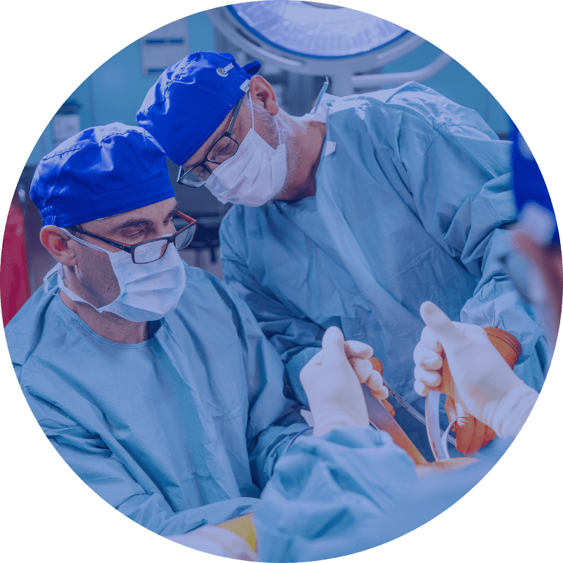
The ACL reconstruction procedure is usually performed under subarachnoid anesthesia. There is no feeling of pain from the waist down. Reconstruction of the anterior cruciate ligament is performed with the use of arthroscopic techniques. In the initial stage of the surgery, the surgeon performs point skin cuts through which a narrow tool ended with a camera is inserted into the joint. Next, through the second point cut, a tool and ligament transplant is inserted, which is fixed in two bone tunnels: femur and tibia.
In this procedure, the damaged ligament is replaced by a thick fascicle, which must be tightened throughout the entire range of the knee joint, and particularly in its last phase, e.g. the ligament. In our hospital we perform reconstructions with the use of various materials. These may be own tendons of the patients, e.g. from the semimembranosus and semitendinosus muscle or from artificial materials. At the end of the procedure, the surgeon rinses the inside of the joint from the blood and leaves the drain in the joint. The rehabilitation of the patient begins the day after surgery. The patient will learn the details of the postoperative period after surgery.
The rehabilitation of the patient begins the day after surgery. Rehabilitation includes individual exercises with a rehabilitator and his help with standing upright for the first time, he teaches how to move with the help of the elbow crutches, as well as gives instructions for individual exercises. The use of CPM braces for continuous passive motion is additional support in the rehabilitation process.