

Surgical treatment of neoplastic and tumour-like lesions of the peripheral nerves and brachial plexus

OPERATIONS
Shoulder arthroscopy Hip endoprosthetics Bunion correction surgery Stem cells ACL reconstruction Knee endoprosthetics Brachial plexus reconstruction Treatment of traumatic peripheral nerve injuries in the extremities Surgical treatment of neoplastic and tumour-like lesions of the peripheral nerves and brachial plexus High density platelet-rich plasma Carpal Tunel release procedure Knee Arthroscopy Hip ArthroscopyOPERATIONS
We can divide tumours of the peripheral nerves into:
(a) originating from the nerve sheath (benign: sheath neuroma, neurofibroma and others, as well as malignant)
(b) originating from nerve cells (neuroblastoma, ganglioneuroma, pheochromocytoma)
(c) tumours metastatic to the nerves
(d) tumours of non-neural origin (lipomatosis, desmoid, lipoma, angioma, intraneural cyst and others)
(e) post-traumatic tumours and those resulting from chronic irritation
Sheath neuroma (schwannoma) is a benign neoplasm, usually occurring between the age of 30 and 60, regardless of the patient’s sex. It usually develops as a single tumour of small to medium size. Within the peripheral nervous system, it usually occurs in the large nerves of the extremities and in the neck (brachial plexus).
Neurofibroma is a benign neoplasm that can be located in large mixed nerves or small branches of sensory nerves.
Malignant peripheral nerve sheath tumours (MPNSTs) can develop in any segment of an originally healthy nerve or originate from a pre-existing benign tumour (mainly neurofibroma, but also schwannoma, ganglioneuroma or pheochromocytoma). MPNSTs can occur in the course of neurofibromatosis type 1 and then, the peak incidence falls within the 3rd decade of life. In the general population, MPNSTs occur in older people with a peak incidence in the 7th decade of life.
The clinical manifestations of a tumour developing intraneurally are associated with compression of the nerve bundles. A palpable tumour is often the first symptom a patient presents to the doctor with. Other symptoms include pain, sensory disorders (weakness, tingling) and movement disorders (muscle atrophy).
Diagnosis of peripheral nerve tumours includes clinical examination and complementary tests (electromyography, X-ray, ultrasound, computed tomography, magnetic resonance imaging, positron emission tomography). In cases of tumours originating in neural tissue, preoperative biopsy of the lesion is not routinely recommended.
Qualification for surgical treatment of peripheral nerve tumours is based on the presence of a palpable tumour mass on clinical examination or its visualization in an imaging test, as well as the coexistence of symptoms such as pain, sensory disorders, motor deficits, positive Tinel-Hoffman sign (tapping the tumour triggers tingling in a specific area of innervation).
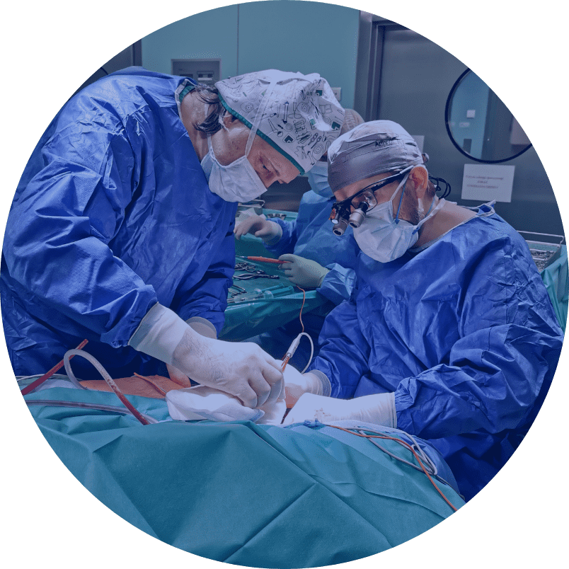
The choice of the suitable surgical technique depends on the intraoperative picture. The presence of a large tumour showing the signs of infiltration into surrounding tissues, with the presence of necrotic foci and extravasation, due to suspected malignancy, is an indication for a biopsy after a longitudinal incision. Benign tumours can be dissected after an incision of the nerve sheath (epineurium). Sheath neuroma resection is possible with little or no disturbance of the bundle structure, since nerve bundles do not pass through the tumour mass. Neurofibroma contains nerve bundles in its composition, so removing the tumour from the nerve requires cutting through these structures. This involves a risk of greater postoperative neurological deficit than after shwannoma resection. Dissecting the tumour from a peripheral nerve requires the use of microsurgical instruments and magnifying optical devices.
Material collected intraoperatively (whole tumour or section) is subject to a histopathological examination, with further management depending on the result.
Recommended length of medical supervision: At least until the results of the histopathological examination; in case of postoperative neurological deficit min. 1 year.
Additional information:
Prognosis: in benign peripheral nerve tumours (they account for approx. 85-90% of the total number of cases) is good, and the risk of recurrence is low. The surgical treatment outcomes depend on the size of the tumour, its location, the nature of the neoplastic histology, the severity of symptoms in the preoperative period and the choice of the suitable microsurgical technique. In most cases, clinical improvement is achieved. In addition, tumour resection prevents further exacerbation of symptoms caused by the tumour mass gradually increasing.
The diagnosis of malignant peripheral nerve sheath tumour (MPNST) requires comprehensive oncological management, while the prognosis in these cases is serious.
Preoperative instructions:
Avoidance of trauma to the tumour area, rehabilitation exercises, electrical stimulation in cases of muscular atrophy, keeping the limb warm.
Postoperative instructions:
Avoidance of overloading the limb in the early postoperative period, ensuring proper wound healing, keeping the limb warm, rehabilitation exercises as recommended.