

Knee endoprosthetics

OPERATIONS
Shoulder arthroscopy Hip endoprosthetics Bunion correction surgery Stem cells ACL reconstruction Knee endoprosthetics Brachial plexus reconstruction Treatment of traumatic peripheral nerve injuries in the extremities Surgical treatment of neoplastic and tumour-like lesions of the peripheral nerves and brachial plexus High density platelet-rich plasma Carpal Tunel release procedure Knee Arthroscopy Hip ArthroscopyOPERATIONS
Knee endoprosthetics surgery is performed primarily due to advanced knee degenerative changes.
Degenerative disease is the most common irreversible joint disease in the case of which articular cartilage is destroyed. In the beginning, the patient feels pain during movement, followed by pains without movement and by night pain. As the disease progresses, there is crackling during movement, limitation of mobility in the joint, sometimes distortion of the limb axis, and at last severe mobility problems.
The rate of disease development varies for each patient. The course of the disease depends on many factors. Common causes of degenerative disease are: obesity, metabolic disorders, systemic diseases, osteochondral necrosis, untreated congenital dislocation of the hip, dysplasia, intraarticular structures damage, inflammatory or post-traumatic changes. In the primary stages of degenerative disease, the doctor may recommend rehabilitation and use of analgesics. Advanced degenerative changes with chronic pain syndrome can be treated only by a procedure of implanting an artificial joint.
A surgical procedure is based on replacing the damaged joint with an artificial one made of wear-resistant materials. Elements forming the artificial joint are the tibia and femoral parts, made of light metal alloy. Between them, there is a polyethylene insert, whose function is to minimize friction.
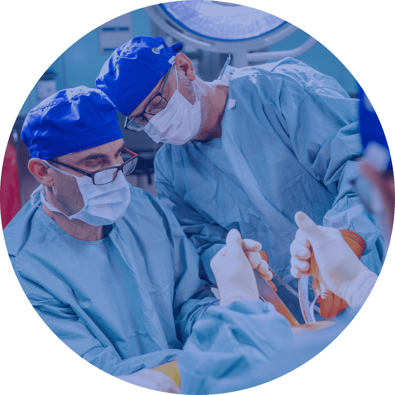
Endoprosthetics is usually performed under subarachnoid anesthesia. There is no feeling of pain from the waist down. Upon reaching the joint, the damaged joint ends of the tibia and thighs are cut. At the next stage of the procedure, the surgeon places a new polyethylene insert between the new artificial joint surfaces. In the final stage of the procedure, it is verified whether all elements fit together and whether the endoprosthesis does not tend to dislocate.
During the first 48 hours after the surgery, a drain is left in the joint. During the first 48 hours after the surgery, a drain is left in the joint. We routinely perform laboratory tests and a test X-ray image in days zero, one and two. The rehabilitation of the patient begins the day after surgery. Rehabilitation consists of individual exercises with the rehabilitant and his aid in the first upright standing, he teaches movement with the help of elbow crutches training as well as gives instructions on unassisted exercises. On the last day the patient learns to move up the stairs. Additional support in the rehabilitation process is the use of CPM braces for continuous passive movement.