

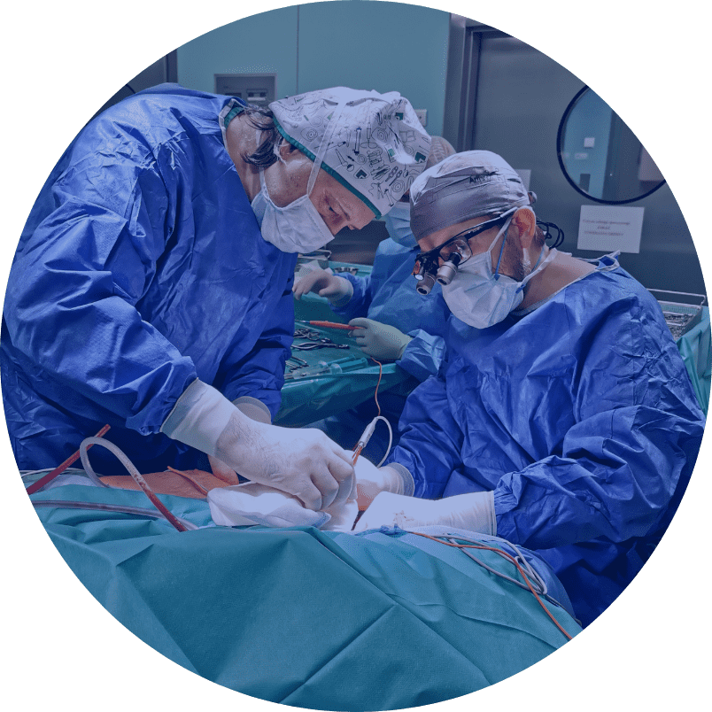
Brachial plexus reconstruction

OPERATIONS
Shoulder arthroscopy Hip endoprosthetics Bunion correction surgery Stem cells ACL reconstruction Knee endoprosthetics Brachial plexus reconstruction Treatment of traumatic peripheral nerve injuries in the extremities Surgical treatment of neoplastic and tumour-like lesions of the peripheral nerves and brachial plexus High density platelet-rich plasma Carpal Tunel release procedure Knee Arthroscopy Hip ArthroscopyOPERATIONS
Damage to the brachial plexus manifests itself as total or partial flaccid paresis [paralysis] of the upper limb and sensory loss. The clinical symptoms that occur depend on the severity of the nerve tissue damage and the type of damage (upper, upper-middle, total).
The most common type is total brachial plexus damage, which manifests itself as a complete inability to perform movements at the humeral joint, elbow and wrist joints, as well as loss of finger and hand grip movements, and lack of surface and deep sensation in the entire upper limb. In addition, muscle atrophy, instability of the shoulder girdle with inferior subluxation of the head of humerus and severe pain gradually exacerbate. In total damage, the upper and middle parts of the brachial plexus may be interrupted or torn out, and the lower part is almost always torn out.
Another type of damage is partial damage, in which the brachial plexus is usually damaged in the upper or upper-middle part, and the lower part is undamaged. In this type of damage, the ability to move the shoulder and elbow joint is lost, while the ability to flex the wrist and fingers is retained. In isolated damage to the upper part, wrist and finger extension is unaffected.
The rarest is damage only to the lower part of the brachial plexus (loss of grasping function of the hand) with preserved function of the upper and middle parts.
The best diagnostic method is clinical evaluation by an experienced brachial plexus surgeon assisted by MRI of the spinal cord at C4-Th2 and nerve conduction test (EMG/ENG). MRI of the brachial plexus or ultrasound are less important in decision making and planning surgical treatment strategies.
During microsurgical brachial plexus reconstructions, we employ the following microsurgical techniques:
– Nerve grafting [implantation] (cable graft) – this involves taking a sensory nerve from another site (usually the sural nerve from the calf or the medial cutaneous nerve of the arm) and using it to fill the defect in the damaged elements of the brachial plexus.
– Nerve transfer – this involves using a healthy motor nerve (e.g., accessory nerve) that innervates a functionally less important muscle and connecting it to the damaged part of the brachial plexus, which innervates the functionally more important muscle. It is also possible to use parts of a healthy nerve, i.e. single bundles.
For the best possible treatment outcomes, we use as many sources of healthy nerves as possible by combining nerve transfer techniques with nerve grafting techniques.
The time between the injury and surgery is of key importance and it affects the outcomes of brachial plexus reconstructive treatment.
The best window for surgery is the first 5 months after injury. Reconstructions performed during this period yield the best results. Operations performed between 6 and 12 months after the injury statistically have worse outcomes. Operations after 12 months from the injury produce unsatisfactory results. In such cases, we no longer perform primary plexus reconstruction; we qualify patients for secondary tenomioplasty or functional muscle graft surgery.
Due to the constant, slow nerve regeneration time of 1 mm/day, we expect the first treatment effects between 7 and 9 months after the surgery (depending on the type of damage and reconstruction strategy), and the final effects approximately 2.5 years after the surgery.
Preoperative instructions:
Maintaining full passive mobility of the limb’s joints, avoiding burns and cuts to innervated parts of the limb, electrical stimulation of the muscles.
Postoperative instructions:
Maintaining immobilization as instructed (on average 4 weeks), then starting rehabilitation exercises, electrical stimulation of muscles after 6 weeks, passive exercises of the limb’s joints, keeping the limb warm, EMG follow-up one year after the surgery and every 6 months thereafter.
- Brachial plexus injury after shoulder dislocation: a literature review. [AUT.] OLGA GUTKOWSKA, JACEK MARTYNKIEWICZ, MACIEJ URBAN, JERZY GOSK. Neurosurg.Rev. 2020 Vol.43 no.2 s.407-423, ryc. tab. bibliogr. 118 poz. summ. DOI: 10.1007/s10143-018-1001-x
- Analysis of patient-dependent and trauma-dependent risk factors for persistent brachial plexus injury after shoulder dislocation. [AUT.] OLGA GUTKOWSKA, JACEK MARTYNKIEWICZ, MAREK STĘPNIEWSKI, JERZY GOSK. BioMed Res.Int. 2018 Vol.2018 art.4512137 [8 s.], tab. bibliogr. 46 poz. summ. DOI: 10.1155/2018/4512137
- Results of operative treatment of brachial plexus injury resulting from shoulder dislocation: a study with a long-term follow-up. [AUT.] OLGA GUTKOWSKA, JACEK MARTYNKIEWICZ, SYLWIA MIZIA, MICHAŁ BĄK, JERZY GOSK. World Neurosurg. 2017 Vol.105 s.623-631, tab. bibliogr. 38 poz. summ. DOI: 10.1016/j.wneu.2017.06.059
- Uszkodzenie splotu ramiennego. [AUT.] JERZY GOSK. W: Chirurgia ręki Warszawa 2017, MediPage Sp. z o.o, s.633-646, ryc. bibliogr. 41 poz, Publikacja w wydawnictwie spoza listy MNiSW, 978-83-64737-84-8.
- The effect of perinatal brachial plexus lesion on upper limb development. [AUT.] JERZY GOSK, WITOLD WNUKIEWICZ, MACIEJ URBAN. BMC Musculoskelet.Disord. 2014 Vol.15 art.116 [7 s.], ryc. tab. bibliogr. 18 poz. summ. DOI: 10.1186/1471-2474-15-116
- Preganglionic injuries in perinatal brachial plexus palsies – results of surgical treatment. [AUT.] JERZY GOSK, ROMAN RUTOWSKI, ROMAN WIĄCEK, MACIEJ URBAN, PIOTR MAZUREK. Neurol.Neurochir.Pol. 2011 Vol.45 nr 2 s.140-147, ryc. tab. bibliogr. 39 poz. streszcz. summ.
- Radiaton-induced brachial plexus neuropathy – aetiopathogenesis, risk factors, differential diagnostics, symptoms and treatment. [AUT.] JERZY GOSK, ROMAN RUTOWSKI, PAWEŁ REICHERT, JERZY RABCZYŃSKI. Folia Neuropathol. 2007 Vol.45 no.1 s.26-30, ryc. bibliogr. 31 poz. summ.
Opracował: prof. dr hab. Jerzy Gosk, dr Jacek Martynkiewicz