

Lumbar microdiscectomy

OPERATIONS
Decompression of lumbar stenosis Discectomy/corpectomy Lumbar microdiscectomy CryoablationOPERATIONS
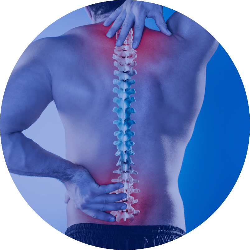
The lumbar microdiscectomy procedure is performed when a symptomatic herniated disc is found in the lumbar spine. Herniation consists of a fragment of the nucleus pulposus of the intervertebral disc and is caused by a rupture of the annulus fibrosus surrounding the nucleus pulposus, causing its fragment to fall outside the natural rim of the intervertebral disc.
This causes compression of the nerve structures in the spinal canal, giving rise to symptoms associated with nerve compression. The most common is low back pain radiating to the lower limb innervated by the compressed nerve. This is also often accompanied by sensory disturbances in the limb (weakened or increased sensation and paraesthesia – a feeling of limb numbness). In some cases, muscle weakness in the limb (paresis in the foot or knee joint) and sphincter dysfunction (inability to urinate or impaired urination control) may additionally occur.
Apart from the characteristic symptoms on neurological examination, the presence of a herniated intervertebral disc and its exact location is determined by magnetic resonance imaging (MRI) of the lumbosacral spine or, if MRI is contraindicated, by computed tomography (CT) of the lumbosacral spine. Operative treatment is indicated if there is no improvement after conservative treatment (rest, pain treatment, rehabilitation) or if there is paresis and/or sphincter dysfunction.
Under general anaesthesia, the patient is laid on the operating table in the prone position on special pads. Using an intraoperative X-ray machine, the intervertebral space where the disc has prolapsed is marked. Then, after disinfecting and draping the surgical site and administering local skin anaesthesia, a linear incision of the skin and subcutaneous tissue, about 3-4 cm long, is made. After reaching the spinous processes of the spine, the muscles are unilaterally (on the side of the prolapsed disc) detached from the bony structures of the spine over a short section.
After reaching the vertebral arches and reconfirming the normal intervertebral space using the X-ray machine in the microscopic image, access to the herniation is performed by making a small bone window and incising the ligamentum flavum, gaining access to the structures of the vertebral canal. The nerve structures of the vertebral canal are identified, primarily the nerve root compressed by the herniated intervertebral disc. After gently dissecting the nerve root under microscopic imaging, the herniation is removed, eliminating the pressure on the nerve structures and ensuring their unobstructed course in the vertebral canal.
Depending on the patient’s individual anatomical features determined intraoperatively, only the fragment of the nucleus pulposus that has prolapsed outside the fibrous ring is removed, or also its fragments inside the ruptured ring. After decompression of the nerve structures, layered closure is performed, and the operation is completed.
Preoperative instructions:
– Up-to-date lumbosacral spine MRI (no more than 6 months old);
– Withdrawal of antiplatelet/anticoagulant drugs under medical supervision of the attending physician (substitute treatment with low-molecular-weight heparin is possible);
– Management of the general condition and adequate treatment of comorbidities, ensuring that the elective surgery under general anaesthesia can be performed.
Postoperative instructions:
– Verticalization and rehabilitation of the patient usually begins during the hospitalisation, the day after surgery;
– Changing dressings, care and observation of the surgical wound, removal of sutures on day 10-12. If fever occurs or the surgical wound appears abnormal, its edges become separated or there is discharge coming out of the wound – contact the hospital immediately;
– Perform exercises as instructed and continue rehabilitation;
– Come for neurosurgical check-up in 1 month and in 3 months;
– Use painkillers as needed.